Influenza A virus Recombinants
by Jo Miles

Abstract
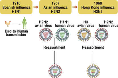
Recombinant reassortment technology was used to prepare H5N1 influenza A virus Recombinants vaccine strains containing a modified hemagglutinin (HA) gene and a neuraminidase gene from isolates A/Hong Kong/156/97 and A/Hong Kong/483/97. and the internal genes of cold attenuated. -adapted influenza A/Ann Arbor/6/60 virus strain. The HA cleavage site (HA1/HA2) of each H5N1 isolate was modified to resemble that of “low pathogenic” avian strains. Five of the 6 basic amino acids at the cleavage site were removed and threonine was added upstream of the remaining arginine.
Modification of the H5 HA cleavage site resulted in the expected trypsin-dependent phenotype without altering the antigenic character of the H5 HA molecule. The temperature-sensitive and cold-adapted phenotype of the original attenuated virus was maintained in the recombinant strains, growing to 108.5–9.4 EID50/mL in eggs. Both H5N1 vaccine virus strains were safe and immunogenic in ferrets and protected chickens against wild-type H5N1 virus challenge.
Viruses and cells
Ca master donor influenza A/AA/6/60 virus (MDV) was obtained from H. Maassab (University of Michigan, Ann Arbor). Influenza A/Duck/Singapore/F 119-3/97 (Dk/Sing/97) is a low pathogenic strain antigenically similar to human isolates from Hong Kong H5N1 provided by the Centers for Disease Control and Prevention (CDC). , Atlanta). MRC-5 and MDBK cells were obtained from the American Type Culture Collection (Rockville, MD). CEK cells, CEF cells, and specific pathogen-free (SPF) embryonated chicken eggs were purchased from SPAFAS (Preston, CT). Chicken antiserum against caMDV was prepared in chicken by Covance (Denver, PA). Ferret antisera against human isolates of H5N1 were prepared at CDC.

Modified HA and wild-type (wt) NA cDNA constructs
Viral RNA (vRNA) was isolated from influenza A/HK/156/97 (wt156) and A/HK/483/97 (wt483) viruses at CDC. Modified HA cDNAs from both viruses were prepared in two fragments by reverse transcription-polymerase chain reaction (RT-PCR) using RNA as a template and two primer pairs: 5′-GCCGCGCGGATC-CTCTTCGAGCAAAAGCAGGGG/5′-TAGTCCTCGAGTCT- CTCTTTGAGG and 5′- GCGAGACTCGAGGACTATTTGGA-G/5′-GCGCGCAAGCTTATTAACCCTCACTAAAAGTGAA-ACAAGGGTGTT.
After digestion with BamHI and XhoI, or XhoI and HindIII, the fragments were ligated into pUC19. The wt NA cDNAs of the wt156 and wt483 viruses were prepared using vRNA as a template and the primers 5′-GCGCGCGAATTCT-CTAGACTCTTCGAGCAAAAGCAGGAGTT and 5′-GCGC-GCAAGCTTATTAACCCTCACTAAAAGTAGAAACAAGG-AGTTT. The NA cDNA was ligated into the EcoRI and HindIII sites of pUC19. Modified HA and NA wt genes were sequenced (373 DNA Sequencer Stretch; Applied Biosystems, Columbia, MD).
Recombinant vaccine candidates
Recombinant viruses containing modified HA and NA wt genes from wt156 and wt483 viruses and the remaining genes from ca A/AA/6/60 virus were prepared using reverse genetics techniques. MRC-5 cells (previously qualified for vaccine production) were used for the transfection of ribonucleoprotein (RNP) complexes derived from the cDNA constructs. In vitro syntheses of HA and NA RNP using the cDNA construct, T3 RNA polymerase, and purified caMDV nucleoprotein were performed as described. RNP was electroporated by a pulse at 320 V and 960 µF into MRC-5 cells 1 h after MDV infection.
HA and NA RNPs were transfected in two separate steps into the background of caMDV. First, the modified HA RNP was transfected into MRC-5 cells that were previously infected with MDV. Tissue culture supernatant was collected 16 h after transfection and passaged twice into chicken eggs in the presence of anti-caMDV antiserum. Transfectants 156HA and 483HA, containing the modified HA gene from wt156 or wt483 virus, respectively, were identified using a hemagglutination inhibition (HI) assay and by RT-PCR analysis.
In addition, the NA RNP of the wt156 virus was transfected into caMDV-infected MRC-5 cells. The supernatant solution was collected 16 h after transfection and used to infect fresh CEK cells. The 156NA transfectant, which contains the NA gene from the wt156 virus in caMDV, was isolated after three passages on CEK cells using limiting dilution. Identification of 156NA was performed by RT-PCR analysis with N1-specific primers.
Analysis of phenotypes and genotypes
The MVS156 and MVS483 phenotypes were verified by plaque assays on CEF cells at 25°C, 33°C, or 39°C. The ca phenotype is defined when the virus titer at 25°C is within 100-fold of the value at 33°C. The temperature-sensitive (ts) phenotype is present when the virus titer at 39°C is at least 100-fold lower than at 33°C. The trypsin dependence of the viruses was tested by plaque assay in CEF and MDBK cells at 33°C. The final concentration of trypsin was 2.5 µg/mL in an agar medium.
The origin of the viral RNA segments in MVS156 and MVS483 was determined by genotype analysis based on the restriction fragment length polymorphism of the PCR product. Each RNA segment was amplified using specific primers. The resulting PCR product was digested with appropriate restriction enzymes. Digestion patterns were compared to wt H5N1 virus and caMDV. The nucleotide sequence of the HA and NA genes was determined (373 DNA Sequencer Stretch; Applied Biosystems, Columbia, MD).

Animal studies
The pathogenicity of MVS156 and MVS483 was determined in the two main SPF chicken varieties available. The first experiment was conducted with White Leghorn chickens at the California Veterinary Diagnostic Laboratory (University of California, Davis). Four groups of 8 4-week-old SPF chickens were inoculated intravenously with 0.2 mL of a 1:10 dilution of the allantoic fluid of each test virus according to US Department of Agriculture guidelines ( USDA).
Inoculated chickens were housed in Horsfal-Bauer units in BSL3+ containment and observed daily for disease or death. On day 10 post-inoculation, serum samples were collected from all surviving chickens and analyzed for seroconversion by HI assay. Five birds from each treatment group were necropsied for histopathological evaluation. Tissues examined included proventriculus, oesophagus, crop, liver, lungs, pancreas, kidney, brain, heart, cecal tonsil, peripheral nerve, and duodenum.
Viruses MVS156 and MVS483 were also tested in White Plymouth Rock chickens at the USDA (Athens GA). A group of 8 SPF chickens were inoculated intravenously (iv) or intranasally and intratracheally (in + it) with 0.2 mL of undiluted test virus allantoic fluid. Chickens were observed daily for disease or death. Serum samples were collected on day 19 after inoculation and treated with a Vibrio cholerae receptor-killing enzyme to determine antibody titers by HI assay. Three weeks after inoculation, surviving chickens from each group were divided into 2 subgroups and challenged in + it with wt156 or wt483 viruses. The chickens were observed for 10 days after exposure for signs of illness or death.
Abstract Recombinant reassortment technology was used to prepare H5N1 influenza A virus Recombinants vaccine strains containing a modified hemagglutinin (HA) gene and a neuraminidase gene from isolates A/Hong Kong/156/97 and A/Hong Kong/483/97. and the internal genes of cold attenuated. -adapted influenza A/Ann Arbor/6/60 virus strain. The HA cleavage site (HA1/HA2) of each H5N1 isolate was…

| M | T | W | T | F | S | S |
|---|---|---|---|---|---|---|
| 1 | 2 | 3 | 4 | 5 | 6 | 7 |
| 8 | 9 | 10 | 11 | 12 | 13 | 14 |
| 15 | 16 | 17 | 18 | 19 | 20 | 21 |
| 22 | 23 | 24 | 25 | 26 | 27 | 28 |
| 29 | 30 | |||||

Categories
- Bird
- Blog
- Conjugation of Synthetic Trisaccharide of Staphylococcus aureus Type 8 Capsular Polysaccharide Elicits Antibodies Recognizing Intact Bacterium.
- Model of vaccine efficacy against HSV-2 superinfection of HSV-1 seropositive mice demonstrates protection by antibodies mediating cellular cytotoxicity.
- My Blog
- PCR
- peptide
- Percp
- Perivascular Lymphocyte Clusters Induced by Gastric Subserous Layer Vaccination Mediate Optimal Immunity against Helicobacter through Facilitating Immune Cell Infiltration and Local Antibody Response.
- peroxidase
- phosphor
- PicoProbe
- Standard
- step
- sterile
- strip
- Target
- TEMED

